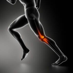
 Chronic exertional compartment syndrome is a nerve and muscle condition induced due to exercise. It leads to pain, swelling and, in some cases, disability in the affected muscles of the arms or legs. While this condition can afflict anyone, it is more common among athletes involved in repetitive impact activities such as running.
Chronic exertional compartment syndrome will often respond to activity modification and non-surgical treatments. However, in a few cases, surgery may be recommended. Most patients can return to their sports or other activities after the surgery. Stellar and sagacious board certified orthopedic surgeons Dr. Steven Thomas and Dr. Gregory Bigler provide treatments for chronic exertional compartment syndrome to patients in Las Vegas, Nevada and surrounding communities in The Silver State.
Diagnosis
Imaging Studies
Imaging tests such as MRI or or near infrared spectroscopy (NIRS) may be performed to diagnose this condition. The muscle structure in the compartments can be assessed with an MRI scan to rule out other possible causes of the symptoms.
An advanced MRI scan may help to evaluate the fluid volumes in the compartments during exercise. This test has been found to be more accurate in identifying compartment syndrome.
NIRS is a new, cutting edge technique that employs the use of light wavelengths to measure the saturation of tissue oxygen in the blood. This helps to determine if the muscle compartment has reduced blood flow.
Compartment Pressure Testing
If an abnormality is not detected with imaging studies, the orthopedic surgeon may recommend measuring the pressure within the muscle compartments. This test is known as compartment pressure measurement, and it is the gold standard for diagnosing chronic exertional compartment syndrome.
The test is mildly painful and invasive, and will involve insertion of needles into the muscles. Therefore, the surgeon will not recommend this test unless the results of other tests and the patient’s medical history strongly suggest the presence of chronic exertional compartment syndrome.
Treatment
Both surgical and non-surgical treatment options are available to treat chronic exertional compartment syndrome. But non-surgical options are usually effective only when the patient quits or significantly reduces the specific activity that is causing the condition.
Non-Surgical Options
The orthopedic surgeon may initially prescribe pain medications, along with a strengthening or stretching exercise regimen, orthotics, massage, a break from regular exercise or sports activity, or the use of various biomechanical techniques, such as altering how to land while jogging. But these non-invasive options are not likely to provide lasting relief from true chronic exertional compartment syndrome.
Surgical Options
The most effective solution for chronic exertional compartment syndrome is surgery. This will involve operating on the inelastic tissue encasing each muscle compartment or fascia. The surgeon may either cut open the fascia of each affected compartment or remove portions of the fascia. The pressure will be relieved with this surgery.
Patients should be aware of the potential surgical risks such as infection, numbness, scarring and permanent nerve damage. Adept and successful board certified orthopedic surgeons at Thomas & Bigler Knee & Shoulder Institute receive patients from Las Vegas, Nevada and other town and suburbs across the landscape for the treatment of chronic exertional compartment syndrome.
If you would like to schedule an appointment or learn more about the Knee and Shoulder Institute procedures & treatments performed by Las Vegas, Nevada board certified surgeons Steven C. Thomas, MD and Gregory T. Bigler, MD. call (702) 933-9393; Physical Therapy (702) 933-9393.
 Cartilage damage can cause stiffness, pain and inflammation in the joint. In case of severe damage, the surgeon will have to perform cartilage repair or removal to restore the joint function. Dedicated and judicious
 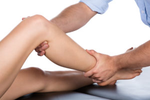 A Baker’s cyst is a fluid-filled cyst that will create a painful bulge and tightness behind the knee. If the patient extends the knee or flexes it fully or is active, the pain may worsen. Also known as a popliteal cyst, it often occurs because of a problem with the knee joint. For instance, a cartilage tear or arthritis can cause the knee to produce excess fluid, resulting in a Baker’s cyst.
 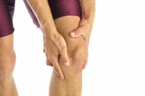 The meniscus is a piece of cartilage that acts as a buffer or cushion between the femur and tibia. Each
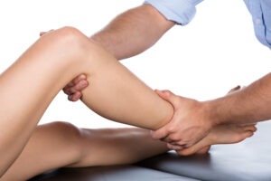
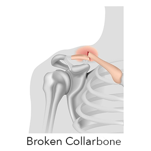 A break in the collarbone is known as a clavicle fracture. Nearly five percent of all adult fractures are clavicle fractures. Most of these fractures can be treated by providing a sling that restricts the movement of the arm and shoulder while the bone heals. But if the bone pieces have shifted to other areas of your body, surgery may be required to realign the collarbone.
 Arthritis of the
 The upper arm bone head rests in a glenoid, which is a shallow socket in the shoulder blade. This head is typically bigger than the socket, and is surrounded by a soft fibrous tissue rim called the labrum. The labrum performs the role of stabilizing the
  ACL or the anterior cruciate ligament runs diagonally in the middle of the knee. A