Describing a Scapula Fracture

Describing a Scapula Fracture

Describing a Scapula FractureA scapula fracture refers to a break in the shoulder blade bone at the back of the shoulder. This type of injury is quite rare, accounting for just one percent of all fractures.

Board certified orthopedic surgeons Dr. Steven Thomas and Dr. Gregory Bigler provide scapula fracture treatments to patients in Las Vegas, Nevada and surrounding locations across the horizon.

Symptoms

Symptoms of a broken shoulder blade would include a sudden pain in the back of the shoulder during the impact as well as rapid inflammation. The patient will feel pain if they attempt to move the upper back or shoulder. The patient will also struggle to lift their arms to their head height. They will feel tenderness, especially over the shoulder blade.

Causes

The patient can experience a scapula fracture or broken shoulder blade if there is a direct impact to the scapula from a blunt article, or due to a fall onto the shoulder or arm. A common cause of this injury is car accidents.

Treatment Approach

Patients should seek prompt medical attention if they fear a scapula fracture. The surgeon may use an x-ray to confirm the fracture and understand how extensive the injury or displacement is. The surgeon will also examine the patient to check for symptoms of vascular or neural injury such as tingling, numbness, weakness or a slower pulse in the arm.

An experienced orthopedic surgeon will treat a scapula fracture conservatively and try to avoid surgery. In this condition, bone displacement is rare as the muscles hold the two (or multiple) parts firmly together.

The surgeon may decide to immobilize the arm in a sling to decrease the arm’s weight on the shoulder. The patient will be recommended to perform early range of motion exercises to maintain movement and avoid stiffness.

A majority of scapular fractures heal in nearly six weeks. After the patient feels pain-free, they can use strengthening exercises restore their total strength and maintain healthy patterns of movement at the shoulder. If the patient’s bone fractures in multiple pieces or there is a significant displacement in the bones, then they may need surgery to fix the scapula back together via pins or wires.

Surgical Treatment

The patient may require surgery if they experience the following kinds of shoulder blade fractures:

  • Fractures of the glenoid articular surface with bone displacement
  • Fractures of the scalpular neck with substantial angulation
  • Fractures of the acromion process that results in the impingement syndrome

During the surgery, the surgeon will initially reposition the bone fragments in their normal alignment.  They will then help the fragments hold together by attaching metal plates with special screws to the outer surface of the bone.

After the surgical procedure, the patient will undergo a period of rehab to enable a safe and healthy recovery of the treated shoulder.

Pain Management

The surgeon will prescribe pain meds for short-term relief after the shoulder blade injury or for a brief duration post-surgery. The surgeon may prescribe a combination of medications such as NSAIDs, Opioids, and local anesthetics for pain management. They will minimize the requirement for opioids as they can become addictive.

Thomas & Bigler Knee and Shoulder Institute, led by board-certified orthopedic surgeons, provides scapula fracture treatments to patients in Las Vegas, Nevada and other towns and cities in The Sagebrush State.

If you would like to schedule an appointment or learn more about the Knee and Shoulder Institute procedures & treatments performed by Las Vegas, Nevada board certified surgeons Steven C. Thomas, MD and Gregory T. Bigler, MD. call (702)

Knowing All About a Supraspinatus Rupture

Knowing All About a Supraspinatus RuptureThe supraspinatus muscle is located along the shoulder blade’s top part and inserts at the top of the humerus bone. It is among the four rotator cuff muscles in the body and is used for lifting the arm up sideways. It is also used significantly for participating in throwing sports because the muscle holds the arm in the shoulder joint when an athlete makes a throw.

After a throwing movement, a strong force is needed to slow down the arm. However, people rarely make an effort to train their supraspinatus muscles. Therefore, a heavy fall on the shoulder can lead to a muscle injury to the tendon.

Outstanding, perspicacious, and board-certified orthopedic surgeons at the Thomas & Bigler Knee & Shoulder Institute provide treatments for supraspinatus fracture to patients in Las Vegas, Nevada, and surrounding communities in The Battle Born State.

Symptoms

A supraspinatus tear is characterized by a sharp shoulder pain during the time of injury. It is accompanied by pain in the arm when it is rotated upward and outward. A patient can also feel more pain and weakness when raising the arm sideways at a 60 degree arc.

Treatment

An athlete who has sustained a supraspinatus injury should apply ice. Ice should not be applied directly on the skin, and should be wrapped in a wet towel to prevent ice burns. A patient can apply ice for 15 minutes every two hours for the first two days after the injury. The frequency for ice therapy can be gradually decreased over a few days. A patient should continue to rest until the pain subsides.

The orthopedic surgeon will usually recommend complete shoulder rest by immobilizing it in a sling, or something similar, for a partial rupture. A patient should consult a sports injury specialist who can advise the best way to go forward with treatment and rehabilitation.

In case of a complete rupture, it may be necessary to undergo a surgery. The surgeon may also recommend an invasive treatment if the conservative approach fails to contain the symptoms.

The success of the surgery will depend on the severity of the injury, the condition of the muscle and the extent of the tear. The surgery will be performed with the help of an arthroscope, which is a device with a miniature camera fixed to it.

The instrument is inserted in the shoulder through a small incision to observe the internal structures to examine the damage. Once the surgeon can identify the cause and extent of damage, they can repair it in a less invasive and more targeted way.

Research studies have shown that supraspinatus ruptures can heal in about 75 percent of the cases. It takes around six months for the patient to fully recover and resume their normal activities. The patient should diligently follow the surgeon’s instructions during both pre- and post-treatment phases in order to achieve safe and sustainable results.

Board certified orthopedic surgeons Dr. Steven Thomas and Dr. Gregory Bigler receive patients from Las Vegas, Nevada, and other suburbs and communities in this part of the country for the treatment of a full or partial suprasinatus rupture.

If you would like to schedule an appointment or learn more about the Knee and Shoulder Institute procedures & treatments performed by Las Vegas, Nevada board certified surgeons Steven C. Thomas, MD and Gregory T. Bigler, MD. call (702) 933-9393; Physical Therapy (702) 933-9393.

Fracture of the Neck of the Humerus

Fracture of the Neck of the HumerusA direct impact on the shoulder or falling onto an outstretched hand can lead to a fractured neck of the humerus. It is a bone that is located at the top of the arm, which forms the glenohumeral joint after fitting into shoulder socket or glenoid.

The humerus neck is at the top of the bone and a break in it results in a fracture, which can happen to an elderly person as well as younger people and children for a variety of reasons. Slipping and falling is a common reason. Tripping over something and hitting the ground with force can cause this injury to occur. Many times children fall off some bars they are playing on or fall out of a tree they are climbing and experience this type of injury.

Honorable, pragmatic, and board-certified orthopedic surgeons at the Thomas & Bigler Knee & Shoulder Institute provide treatments for humerus fracture to patients in Las Vegas, Nevada, and surrounding locations in The Sagebrush State.

Treatment

A fracture that has more than two fragments, such as those with a displacement over 1 cm or leading to shoulder dislocation usually requires a surgery.

An orthopedic surgeon will treat a displaced fracture more conservatively, and try to avoid surgery. Such fractures heal quickly and the patient is asked to wear a broad arm sling. They are best treated with a cuff and collar that allows gravity to rectify any angulation or misalignment.

The surgeon will advise the patient to keep the arm, which is in a sling, underneath the clothes for extra protection for the first two weeks. After two weeks, the patient will be prescribed shoulder joint pendular exercises.

From the fourth week onwards, the patient is allowed to wear the cuff and collar outside the clothes. Gradually, as the arm improves over the next two weeks, the cuff and collar are taken off.

Non-Surgical Treatment

Immobilization

The patient is made to wear a sling to provide arm immobilization and support. Another option is to use a swathe, which offers added support and reduces movement further as it is wrapped horizontally around the arm and chest.

The surgeon will usually prescribe arm immobilization for around two to three weeks. But the duration can vary depending on the patient’s medical condition, age, and fracture type.

Pain Management

A patient will be prescribed pain medications and anti-inflammatory drugs like ibuprofen during the healing process.

Range of Motion Exercises

The patient will be required to perform specific ROM exercises, but only after 7 to 10 days of sustaining the injury.

Physical Therapy

The surgeon may recommend physical therapy after 2 to 3 weeks of the injury.

Surgical Treatment

A broken proximal humerus will have to be surgically replaced or repaired. A surgeon may secure the bone pieces together with the help of screws, plates, and nails. If the surgery cannot repair the impacted bone, then the shoulder ball may have to be replaced during the surgery.

Surgery is usually recommended for fractures that involve rotator cuff attachments, displaced fractures in younger patients, cases where there is a head-split fracture in the humerus, injuries involving nerves and blood vessels, open fractures, or dislocated fractures.Awesome board certified orthopedic surgeons Dr. Steven Thomas and Dr. Gregory Bigler receive patients from Las Vegas, Nevada, and other towns and suburbs in this part of the state for the treatment of a fractured neck of the humerus.

If you would like to schedule an appointment or learn more about the Knee and Shoulder Institute procedures & treatments performed by Las Vegas, Nevada board certified surgeons Steven C. Thomas, MD and Gregory T. Bigler, MD. call (702) 933-9393; Physical Therapy (702) 933-9393.

Pec Major Tendon Strain (Rupture)

Defining a Pec Major Tendon Strain (Rupture)The pectoralis muscle, which is located at the front of the chest, is a large and strong muscle that rotates the arm inward. The muscle’s weak point is the tendon where it is attached to the arm bone. It is the point that is likely to get ruptured during weight training activities, such as a bench press.

Cordial, innovative, and board certified orthopedic surgeons at the Thomas & Bigler Knee & Shoulder Institute provide treatments for pec major tendon strain or rupture to patients in Las Vegas, Nevada, and surrounding locations across the landscape.

Symptoms

A pec major tendon sprain is characterized by a sudden and sharp pain at the upper arm’s front part, near the shoulder, where the pec major tendon is attached. The strain is accompanied by a rapid swelling of the upper arm and the front of the shoulder.

An orthopedic surgeon will diagnose the injury by reproducing the pain to confirm the cause. It is done by making the patient pull their arm across the front of the chest or rotate the arm inward against resistance. There is also a noticeable lump or gap in the muscle.

Treatment

An athlete who gets a pec major tendon sprain should follow the principle of R.I.C.E, which is rest, ice, compression, and elevation, for a minimum of two days after the injury. The patient should apply ice for 10 to 15 minutes every hour and decrease the application as the symptoms gradually diminish.

A patient should also consult a sports injury specialist. If the tendon is fully ruptured, then the surgeon will recommend a surgery, followed by a long duration of rest and a subsequent full rehabilitation program. Applying a sports massage to the injured area can also help in healing the injury after the acute stage.

Repairing a Torn Pectoral Tendon

Patients undergoing a surgery to repair the pec tendon tear should how it may be repaired.

Type 1 Tear

It is simple to repair these kinds of tears where the tendon is torn off the bone. It involves an open surgery where an incision is placed into the patient’s axilla.

Surgeons will use a suture anchor device during the open surgery to fit the sutures into the bone, which will then be used to attach the tendon back to the bone. The patient will be in a sling for a relatively longer duration to ensure enough healing before starting on strengthening exercises.

Type 2 Tear

A surgeon will wait until some early repair scar tissue has formed on the pec muscle because this makes a rather difficult repair easier to carry out. Surgeons can sometimes use the cadaver tendon to strengthen or augment the repair.

Type 3 Tear

These tears occur rarely, and mostly within the muscle. They are hard to repair and the surgeon may wait for a couple of weeks so that scar tissue can form and facilitate the repair process.

Terrific, perceptive, and board certified orthopedic surgeons Dr. Steven Thomas and Dr. Gregory Bigler receive patients from Las Vegas, Nevada, and other neighborhoods and cities in this region of the southwest for the treatment of pec major tendon strain or rupture.

If you would like to schedule an appointment or learn more about the Knee and Shoulder Institute procedures & treatments performed by Las Vegas, Nevada board certified surgeons Steven C. Thomas, MD and Gregory T. Bigler, MD. call (702) 933-9393; Physical Therapy (702) 933-9393.

Long Head of Biceps Rupture on the Shoulder

Long Head of Biceps Rupture

The biceps muscle bifurcates into two tendons at the shoulder, a long one and a short one. The long tendon lies over the top of the humerus bone and attaches right at the top of the shoulder blade. It can be impacted by a partial rupture that leads to pain at the shoulder front.

Board certified orthopedic surgeons at the Thomas & Bigler Knee & Shoulder Institute provide treatments for biceps rupture to patients in Las Vegas, Nevada, and other towns and communities in this section of Nevada.

Symptoms

Long head of biceps rupture is characterized by a sudden sharp pain at the upper arm’s front part, which can also be accompanied by swelling and pain over the front portion of the shoulder joint. A patient can find it painful to contract the biceps muscle against resistance, particularly in the first few days when it becomes difficult to lift the long head of the biceps, straight up forward over the head, and against resistance.

Treatment

Athletes who may have ruptured the long head of the biceps should apply ice on the injured area or use cold therapy as emergency first aid. The patient should also immediately consult a sports injury specialist. It is vital to take adequate rest, and not make too many movements if the patient is in pain.

An orthopedic surgeon will prescribe treatment followed by a detailed rehabilitation program. A patient may also require surgery to repair the tendon, if there is a severe or complete rupture.

Rehabilitation Program

The injured biceps tendon can benefit from occupational or physical therapy based on the individual case, surgeon’s recommendation and the type of facility. The surgeon will advise rehabilitation after a complete examination of the area to understand if there are any associated injuries and complicated factors that may impact the treatment.

They will ask the patient to rest the impacted joint in the acute stage, which can be also be accompanied by soft immobilization. The therapy may also include controlling the swelling with cold therapy, such as using specialized cold compression units, hydrotherapy, ice massage, and cold packs. The inflammation can be treated with non-steroidal anti-inflammatory drugs.

A surgeon will advise post-op rehabilitation after examining the intra-operative findings during repair. The surgeon may put the patient’s arm in a soft sling immediately after the surgery, which will allow the patient to make gentle movements of the arm and range of motion (ROM).

To initiate ROM and muscle strengthening, the surgeon can introduce therapy bands and pulleys after 10 to 14 days of the surgery. The patient is advised to do advanced functional exercises as well as maintain and advanced ROM for six to eight weeks.

During this time, the affected arm can tolerate most cases of moderate loading. However, the patient is advised against heavy loading for a few more months, particularly in case of distal tendon repairs.

Profound, kind, and board certified orthopedic surgeons Dr. Steven Thomas and Dr. Gregory Bigler receive patients from Las Vegas, Nevada, and other parts of this region of the state for the treatment of long head of biceps rupture.

If you would like to schedule an appointment or learn more about the Knee and Shoulder Institute procedures & treatments performed by Las Vegas, Nevada board certified surgeons Steven C. Thomas, MD and Gregory T. Bigler, MD. call (702) 933-9393; Physical Therapy (702) 933-9393.

Shoulder Sprain

Shoulder Sprain | Las Vegas Orthopedic Surgery | Orthopedic Surgeons
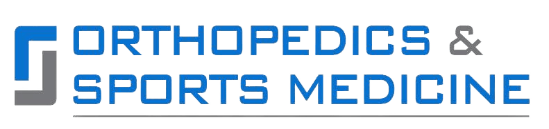
Shoulder Sprain Treatment

A shoulder sprain occurs when there is a tear in the ligaments, which are strong bands of fibrous tissues connecting one bone to another inside or around the joint of the shoulder. It is a common misconception that the shoulder has a single joint between the humerus and the torso, though it actually has numerous smaller joints outside the socket of the arm bone.

Judicious and board certified orthopedic surgeons at the Thomas & Bigler Knee & Shoulder Institute provide treatments for shoulder sprain to patients in Las Vegas, Nevada, and surrounding locations across the horizon.


Types of Shoulder Sprain

Spraining the shoulder could indicate a fiber stretch as well as a partial or a fully torn ligament or joint capsule. A ligament tearing sprain in the shoulder mostly takes place at the joint between the collarbone and acromion, referred to as the acromioclavicular joint.

A shoulder sprain is sometimes also known as a shoulder separation. In certain cases, a shoulder sprain may occur in the sternoclavicular joint between the collarbone and the breastbone, which is located within an inch of the chest’s midline.


Symptoms

The symptoms of shoulder sprain vary according to how serious the injury is. It can range from mild to severe, and includes shoulder pain, usually at the joint’s front portion. A patient can also feel tenderness if they press the injured area. Really bad shoulder sprains can lead to a shoulder joint instability.


Causes

A force or a heavy impact on the arm, which stretches the shoulder ligaments, usually causes a sprained shoulder. This occurs particularly when the force makes the arm go back when it is raised to a level of 90 degrees at the shoulder. The impact can stretch or tear the capsule or ligaments at the front of the shoulder.

A shoulder sprain is not a common occurrence because the shoulder ligaments are generally quite strong. Moreover, the muscles located at the shoulder’s front portion, such as the pectorals, are more inclined to get injured first.


Treatment

The sprain type and its grade influence the injury’s treatment.

Acromioclavicular joint sprains

If it is a Grade I or II sprain, then the surgeon will advise rest, cold therapy, and a non-steroidal anti-inflammatory drug such as ibuprofen to get relief from swelling and pain. The arm is also placed in a sling for one to three weeks. If the injury is of Grade III, then the sling has to be worn for four weeks, and in some cases, surgery may be required.


Sternoclavicular joint sprains

For Grade I sprains, rest, cold therapy and non-steroidal anti-inflammatory drugs are prescribed. The arm is put in a sling for one to two weeks. If the injury is of Grade II level, then the patient has to wear the sling for three to six weeks. Grade III sprains are treated with a procedure called closed reduction, where the displaced collarbone is carefully put back into place.

Outstanding, stellar, and board certified orthopedic surgeons Dr. Steven Thomas and Dr. Gregory Bigler receive patients from Las Vegas, Nevada, and other towns and cities in this part of America for the treatment of shoulder sprains.

If you would like to schedule an appointment or learn more about the Knee and Shoulder Institute procedures & treatments performed by Las Vegas, Nevada board certified surgeons Steven C. Thomas, MD and Gregory T. Bigler, MD. call (702) 933-9393; Physical Therapy (702) 933-9393.

Knowing All About Rotator Cuff Tendonitis

Rotator cuff tendonitis, also known as rotator cuff tendinopathy, is a degenerative condition that affects one or more of the rotator cuff tendons located on the shoulder. A previous injury such as a rotator cuff partial dislocation or strain can lead to rotator cuff tendonitis. 

The condition is one of the most common causes of shoulder pain. A patient can feel it coming on gradually over time or after straining the rotator cuff because it has not healed properly.

Terrific and board certified orthopedic surgeons at the Thomas & Bigler Knee & Shoulder Institute provide treatments for rotator cuff tendonitis to patients in Las Vegas, Nevada, and surrounding locations across the horizon.

Symptoms

Shoulder tendonitis is associated with shoulder pain while resting or due to certain movements. The pain may occur even while sleeping at night. A patient also feels pain when performing overhead activities such as playing racket sports or swimming.

However, pain caused by arm movements below shoulder height is less common in rotator cuff tendinopathy. A patient may also feel a creaking, referred to as crepitus, while performing shoulder movements.

Treatment

The condition can be diagnosed and any tearing of the tendon can be identified with the help of a MRI scan. The rotator cuff tendinopathy treatment includes two stages. The first part consists of treating the symptoms to reduce the inflammation and pain, so that the patient can make normal movement. The second stage includes addressing the underlying causes of the condition and correcting them.

As a first part of the treatment, the patient should perform RICE therapy as follows:


  • Rest: The patient should refrain from activities that cause pain. Rest is advised.


  • Ice or cold therapy: Pain and inflammation can be reduced with the help of ice or cold therapy. The patient can apply ice for 10 minutes every hour. The frequency can be gradually reduced to applying ice for 15 minutes every three to four hours, based on the state of the condition.


  • Exercises: It is imperative for patients to exercise once the pain and inflammation has subsided enough to allow it.

Other Treatments

Medication

The surgeon may prescribe ibuprofen or other anti inflammatory medication that brings about pain relief in the early stages of the injury.

Electrotherapy

Treatments such as magnetic field, laser, interferential stimulation, or ultrasound may be helpful in mitigating pain and inflammation.

Massage

A sports massage may bring about relaxation and loosen the tightness of the shoulder muscles. A cross friction massage on the tendon can be helpful if cold therapy or other initial conservative treatment cannot address the chronic injury.

Corticosteroid Injections

Injecting corticosteroid into the subacromial space of the shoulder can help reduce the pain and inflammation as well as facilitate the initiation to an exercise rehabilitation program.

Treatment may be difficult if there is tendon calcification or bone growth. In such cases, the surgeon may suggest keyhole surgery. Fantastic and board certified orthopedic surgeons Dr. Steven Thomas and Dr. Gregory Bigler receive patients from Las Vegas, Nevada, and other suburbs and neighborhoods in The Silver State for the poignant treatment of shoulder tendonitis.

If you would like to schedule an appointment or learn more about the Knee and Shoulder Institute procedures & treatments performed by Las Vegas, Nevada board certified surgeons Steven C. Thomas, MD and Gregory T. Bigler, MD. call (702) 933-9393; Physical Therapy (702) 933-9393.

Posterior Shoulder Dislocation

A posterior shoulder dislocation takes place when the head of the long bone in the upper arm, known as the humerus, moves backward out of the socket. The humerus bone is most likely felt at the back of the joint. It is quite a rare shoulder injury because the majority of shoulder dislocations are anterior. Proximal Humerus Growth Plate Fracture | Las Vegas Orthopedic Surgery

Remarkable, sagacious, and board certified orthopedic surgeons at the Thomas & Bigler Knee & Shoulder Institute provide treatments for posterior shoulder dislocation to patients in Las Vegas, Nevada, and surrounding towns and neighborhoods in this part of the US.

Symptoms

The symptoms of a dislocated shoulder posterior include excruciating pain in the joint of the shoulder at the time of injury, followed by rapid swelling. The condition leads to a total loss of shoulder function and the patient is not only in constant pain but is also unable to use the arm. The afflicted shoulder also has a deformed appearance and the patient usually wants to hold out the arm to the side and rotate it inward.

Causes

Shoulder posterior dislocations usually happen due to two reasons, either because of a direct hit to the humeral head’s front, or by falling onto an outstretched arm. Both the causes leading to shoulder posterior dislocations can occur due to sports activities, particularly contact sports. These may also occur due to trauma caused by vehicle accidents and epileptic seizures where the patient violently thrashes their arm.

Treatment

A patient should seek medical attention immediately for posterior shoulder dislocation. They should not move their shoulder or try to put it back into position on their own. An orthopedic surgeon will check the hand, wrist, sensations and pulses in the arm to determine if there is any blood vessel or nerve damage.

For minor dislocations and conditions where there are no associated fractures, a surgeon can actually pop back the dislocated bone back into place. In any case the patient will be administered an anesthetic, sedatives, or relaxants before such a step is carried out.

The surgeon will then prescribe adequate rest to give time to the soft tissues in the shoulder to heal. Subsequently, the patient will be put on a rehabilitation program to regain strength and full movement.

Severe cases that involve fractures, damage to nerves or blood vessels, or extensive soft tissue injury may require surgery to treat the humeral head and repair the injured structures. Surgery will have to be followed by a period of complete rest and a comprehensive rehabilitation program.

Surgery

A surgeon may operate when the patient has recurrent dislocation or subluxation. In such cases, an individual’s torn cartilage and ligaments are reattached to the bone with stitches or sutures.

If the subluxation or dislocation is secondary to the stretched ligaments, then first the surgeon will tighten the ligament with a process called capsular shift or capsulorraphy, and then secure it with sutures or stitches.

If a patient has stretched ligaments, which are also torn away from the bone, then both the procedures can be carried out. Golden and board certified orthopedic surgeons Dr. Steven Thomas and Dr. Gregory Bigler receive patients from Las Vegas, Nevada, and nearby areas in The Battle Born State for the treatment of posterior shoulder dislocation.

If you would like to schedule an appointment or learn more about the Knee and Shoulder Institute procedures & treatments performed by Las Vegas, Nevada board certified surgeons Steven C. Thomas, MD and Gregory T. Bigler, MD. call (702) 933-9393; Physical Therapy (702) 933-9393.

Long Head of Biceps Inflammation

The biceps muscle bifurcates into two tendons at the shoulder, which is the long tendon and the short tendon. The long tendon goes over the upper arm’s top and attaches to the shoulder blade’s top.

Long tendon inflammation of the biceps muscle, also known as biceps tendinitis, is a common condition that may typically affect weightlifters, golfers, throwers, rowers, and swimmers.

Innovative, cordial, and board certified orthopedic surgeons at the Thomas & Bigler Knee & Shoulder Institute provide treatments for biceps inflammation to patients in Las Vegas, Nevada, and other cities and suburbs in The Sagebrush State.

Symptoms

Biceps tendon inflammation is characterized by pain at the shoulder’s front where the biceps muscle’s long head attaches. A patient can isolate the biceps tendon by lifting their arm straight up in front against resistance, which reproduces the pain at the shoulder front. An individual can also feel tenderness when they press over the tendon at the same point.

Treatment

An athlete inflicted with long head of biceps inflammation should rest until the pain subsides. The patient should apply ice to get relief from pain and inflammation. The patient should apply ice for 10 to 15 minutes every hour in the beginning and thereby should reduce the frequency as there is gradual improvement. Patients must also consult a sports injury professional.

A sports injury specialist will prescribe ibuprofen or other anti-inflammatory medication. The specialist or a physical therapist may also demonstrate strengthening and stretching exercises as a part of a full rehabilitation program. A trained theapist may give a sports massage to the injured muscle. If the case is chronic then a sports injury specialist can apply a cross friction massage to the tendon.

Non-Surgical Treatment

An orthopedic surgeon will usually start the treatment of biceps tendinitis with simple methods, such as prescribing non-steroidal anti-inflammatory medicines and drugs like naproxen and ibuprofen to reduce swelling and pain. The surgeon may also prescribe cortisone steroids, which are impactful anti-inflammatory medicines. Steroid injections in the tendon can bring about pain relief.

Physical therapy, including specific strengthening and stretching exercises may also be prescribed to help restore the strength and motion of the shoulder.

Surgical Treatment

If the patient’s condition does not become better with non-surgical treatment, then a surgeon may offer to operate. Surgery also makes for a prudent option if the patient is afflicted with other shoulder problems.

Arthroscopic Surgery

Arthroscopic surgery involves inserting of an arthroscope and very small surgical instruments into the joint of the shoulder. Biceps tendinitis surgery is generally performed arthroscopically as it allows the surgeon to assess the biceps tendon’s condition as well as the other shoulder structures.

The arthroscope is a small camera that shows images on a television screen, which are used by the surgeon to guide the miniature surgical instruments.

Biceps Tenodesis

In some cases, a surgeon can carry out biceps tenodesis where the bicep’s damaged section is removed and the remaining tendon is reattached to the humerus. Salient, astute, and board certified orthopedic surgeons Dr. Steven Thomas and Dr. Gregory Bigler receive patients from Las Vegas, Nevada, and other neighborhoods and towns in this area of the southwest for the treatment of biceps inflammation.

If you would like to schedule an appointment or learn more about the Knee and Shoulder Institute procedures & treatments performed by Las Vegas, Nevada board certified surgeons Steven C. Thomas, MD and Gregory T. Bigler, MD. call (702) 933-9393; Physical Therapy (702) 933-9393.

Clavicle Muscle Attachments Inflammation

What is Clavicle Muscle Attachments Inflammation?

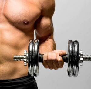
The collarbone, medically known as the human clavicle, is a short bone that connects the arms to the trunk. One can easily feel it by placing their hand on the area of the skin, which is right above the first rib. The clavicle can be easily seen in thin people.

The human clavicle is a resilient and strong bone that takes the weight of the upper limb, while remaining firmly in its position to give stability to the muscles as well as the ligaments attached to it.

Board certified orthopedic surgeons at Thomas & Bigler Knee and Shoulder Institute provide treatments for clavicle muscle attachments inflammation to patients in Las Vegas, Nevada, and surrounding communities.

Swollen Clavicle

Any type of enlargement near the collarbone or fluid accumulation can lead to a swollen clavicle. In certain cases, the swelling is not related to the collarbone but rather a soft tissue injury that commonly impacts the muscles and subcutaneous tissue, which further leads to swelling over the collarbone.

The swelling could be due to any of the muscles that attach the collarbone or the platysma muscle, an area right over the clavicle. Clavicle swelling is also referred to as bony swelling and is more noticeable with a fracture.

Causes of Swollen Clavicle

Any of the following reasons can cause a swollen clavicle:

  • Injury: A bone injury or a trauma in the surrounding tissue can lead to a swollen clavicle.
  • Bone disorders: Certain bone growth disorders like Paget’s disease can lead to a swollen collarbone.
  • Infection: A clavicle infection can lead to pain and swelling in the surrounding tissue; however, the chances of this occurring are rare.
  • Vitamin D deficiency: Inadequate intake of vitamin D can decrease the amount of calcium the body gets, which can also result in bone deformation and a swollen clavicle.
  • Arthritis and osteoarthritis: These afflictions can produce a wear and tear effect on joints, resulting in a swollen collarbone.
  • Joint problems: An inflammation of the joints surrounding the collarbone can also lead to a swelling of the clavicle.

Treatment for Swollen Clavicle

One should treat a swollen clavicle according to the underlying cause. For instance, a swollen clavicle caused by fracture or injury should be treated by keeping the arms immobile with the help of a sling.

If arthritis is the cause of a swollen clavicle, then the most common treatments are rest and taking pain medication. However, one might also need physiotherapy or surgery depending on the seriousness of the condition.

In case of infection-related swollen clavicle, it is necessary to treat the underlying infection to address the swollen lymph nodes.

Patients should consult with an orthopedic surgeon immediately if they notice tenderness, redness, or pain associated with the swelling. A swollen clavicle can quickly return back to its normal state with adequate diagnosis and treatment.

Board certified orthopedic surgeons Dr. Steven Thomas and Dr. Gregory Bigler receive patients from Las Vegas, Nevada, and nearby areas for clavicle muscle inflammation treatment.

If you would like to schedule an appointment or learn more about the Knee and Shoulder Institute procedures & treatments performed by Las Vegas, Nevada board certified surgeons Steven C. Thomas, MD and Gregory T. Bigler, MD. call (702) 933-9393; Physical Therapy (702) 933-9393.